

Valvular heart disease
Valvular heart disease represents a significant challenge in the field of cardiovascular medicine. They include various conditions such as insufficiencies and stenoses, which can often have serious effects on heart function. Through advances in research and the development of innovative interventional therapies, the treatment of these conditions has evolved significantly. Nevertheless, they remain a common cause of morbidity and mortality and require a multidisciplinary approach for optimal outcomes.
Ongoing Projects
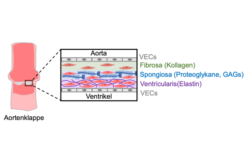
Source: AG Sengle, Cologne
The biomechanical function of heart valves is determined by the extracellular matrix (ECM) of the valve’s own cells, such as vascular endothelial cells (VECs) and vascular interstitial cells (VICs). The ECM supports the structure and function of the valves and regulates the bioavailability of signaling molecules. In healthy valves, the ECM is divided into three layers: the collagen-rich fibrosa, which provides strength; the elastin-containing ventricularis, which provides flexibility; and the proteoglycan-rich spongiosa, which cushions movement.
Clinically significant heart valve diseases often result from disturbances in the ECM structure caused by genetic, mechanical, or inflammatory factors. Congenital heart valve diseases, such as the bicuspid aortic valve, as well as acquired conditions, such as calcification and myxomatous degeneration, are common. Risk factors such as male gender, high blood pressure, and smoking increase the strain and wear on the valves over the course of a lifetime. Inflammatory mechanisms also play a role.
The goal of our research is to understand how defects in ECM architecture influence the development and progression of heart valve diseases.
Our primary goals are:
- To gain a better understanding of the ECM architecture required for proper heart valve function.
- To understand how the microenvironment controls the behavior of resident cells and immune cells.
- To develop therapeutic strategies to counteract valvular diseases by blocking harmful “outside-in” signaling pathways to VECs and VICs.
Sengle Lab - Cologne
Research Group on Congenital Connective Tissue Disorders, Fibrillinopathies
Publications
Carmen Yap, Myrthe E. Hoogeland, Siyu Li, Iris Bakker, Ela Yalcin, Roelof Ottenhoff, Maria P. Clemente-Olivo, Sander Kooijman, Yousef Morcos, Gerhard Sengle, Vivian de Waard,
A high-fat diet does not exacerbate aortic pathology and enhances metabolic function in Marfan syndrome Fbn1C1041G/+ mice,
Biochimica et Biophysica Acta (BBA) - Molecular Basis of Disease, Volume 1872, Issue 3, 2026, 168107, ISSN 0925-4439, doi.org/10.1016/j.bbadis.2025.168107.
de Souza, R.B., Cassiano, L.L., Barnowski, P., et al. Exploring thoracic aorta ECM alterations in Marfan syndrome: insights into aorta wall structure. Sci Rep 15, 26665 (2025). https://doi.org/10.1038/s41598-025-09665-w
Spanou CES, Yang C, Godwin ARF, et al. Prodomain processing controls BMP-10 bioactivity and targeting to fibrillin-1 in latent conformation. The FASEB Journal. 2025; 39:e70373. doi:10.1096/fj.202401694R
Spanou CES, Wohl AP, Doherr S, et al. Targeting of bone morphogenetic protein complexes to heparin/heparan sulfate glycosaminoglycans in bioactive conformation. The FASEB Journal. 2023; 37:e22717. doi:10.1096/fj.202200904R
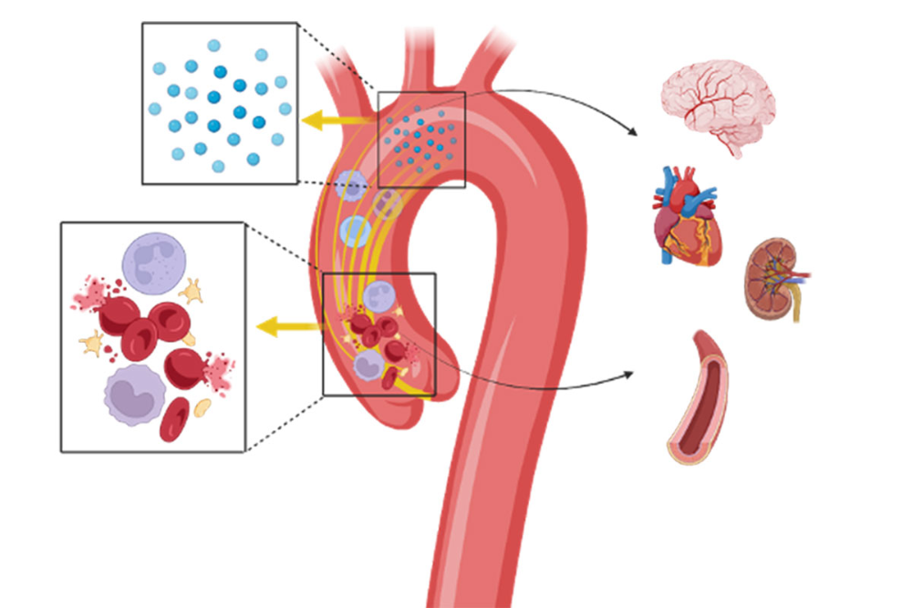
Hemodynamic forces and biomechanical properties determine the conductivity and vulnerability of the aortic wall, but the exact mechanisms remain unknown. This project uses magnetic resonance imaging (MRI) for regional hemodynamic mapping of the aortic valve as well as the thoracic and abdominal aorta. The goal is to identify patterns in the development of aortic diseases, including aneurysms and aortic valve stenosis.
A combined MRI approach (4D flow, DTI, X-nucleus MRI) will evaluate hemodynamic parameters, vascular changes, and cellular profiles in the same region. This is done in the context of myocardial function and structural and metabolic changes using cardiovascular magnetic resonance imaging (CMR). Repeated regional phenotyping will compare novel MRI metrics throughout the course of aortic diseases, including in patients who have undergone aortic valve replacement.
Pathological flow patterns, particularly in the ascending aorta, are characterized by increased velocity, turbulence, and inhomogeneous shear stress. These factors affect circulating blood components and lead to organ damage. Aortic valve stenosis (AVS) causes endothelial dysfunction, which can be improved by valve replacement. This underscores the importance of regulated blood flow in the aorta for cardiovascular health.
- Hybrid Imaging in Structural Interventions
- Effect of aortic valve stenosis on the function of the supravalvular aorta
- Myocardial adaptation in aortic valve stenosis
- Physicomechanics of the aorta and systemic vascular function—diagnostics and monitoring
- CD40 Signaling in Aortopathies
Quast Lab, Gerdes Lab, Jung Lab, Zeus Lab, Nienhaus Lab – Düsseldorf
Quast Research Group – University Hospital Düsseldorf
Cardiovascular Immune Biology – PI Gerdes – University Hospital Düsseldorf
Jung Research Group – University Hospital Düsseldorf
Publications
Isabella Solga, Leon Götzmann, Vithya Yogathasan, Patricia Wischmann, Tin Yau Pang, Patricia Kleimann, Sebastian Temme, Lina Hofer, Anja Stefanski, Alexander Lang, Georg Nickenig, Christian Jung, Norbert Gerdes, Mohammed Rabiul Hosen, Malte Kelm, Ramesh Chennupati, “Large extracellular vesicles derived from red blood cells in coronary artery disease patients with anemia promote endothelial dysfunction,” Journal of Molecular and Cellular Cardiology, 217, (76–88), (2026).https://doi.org/10.1016/j.yjmcc.2026.06.010
Calcific aortic valve stenosis (AVS) is the most common heart valve disease in the aging population. To date, there is no drug therapy available. The only treatment is surgical or interventional valve replacement. A key driver of the disease is sterile inflammation of the valve—that is, inflammation not triggered by pathogens. At the center of this inflammatory response is the NLRP3/ASC inflammasome, a molecular complex of innate immune cells.
Upon activation of the inflammasome, the adapter protein ASC forms large aggregates known as “ASC specks.” These are released into the extracellular space, where they sustain or exacerbate the inflammation. We are investigating the hypothesis that extracellular ASC specks are a key driver of chronic valve inflammation and calcification.
In this project, funded by the German Research Foundation (DFG), we are developing and characterizing nanobodies (VHH, single-domain antibodies from camelids) that specifically bind to and neutralize ASC specks. The goal is to interrupt inflammation-driven calcification at a defined molecular target, as a potential first pharmacological treatment approach for ACS. The project is being carried out in close collaboration with the Institute for Innate Immunity (Franklin Group) and the Nanobody Core Facility in Bonn.
Our primary objectives are:
- To characterize extracellular ASC specks as drivers of valve calcification.
- To develop and validate ASC-specific nanobodies (VHH^ASC).
- To test the nanobody-based therapy in preclinical models of aortic valve stenosis.
- Ultimately, to establish a pharmacological treatment for AVS, which to date has only been treatable surgically or through interventional procedures.
Franklin Lab, Bartsch Lab – Bonn
The research group is dedicated to basic cardiovascular research with a particular focus on inflammatory diseases such as atherosclerosis and aortic valve stenosis. The goal of the research is to better understand the molecular mechanisms underlying these diseases and to identify new therapeutic targets.
To this end, modern cell and animal models are used to discover potential target structures and investigate their relevance in the progression of the disease. The results are then validated in human cells and patient samples to assess their clinical relevance and pave the way for potential therapeutic translation.
This interdisciplinary approach combines experimental basic research with translational medicine—with the goal of developing innovative therapeutic approaches for cardiovascular diseases and thus contributing to the long-term improvement of patient care.
Our research focuses on the therapeutic utilization of brown algae and the identification of therapeutic targets in the endocannabinoid system.
Our work is funded by the DFG, the BMFTR, and the EIC.